[最も共有された! √] e coli cell under microscope 341289-How does e coli look under a microscope
The length can range from 110 µm for filamentous or rodshaped bacteria The most wellknown bacteria E coli, their average size is ~15 µm in diameter and 26 µm in length In this figure The size comparison between our hair (~ 60 µm) and E coli (~1 µm) Notice how small the bacteria areMETHODOLOGY Open Access Reliable measurement of E coli single cell fluorescence distribution using a standard microscope setup Marilisa Cortesi1*, Lucia Bandiera1,6, Alice Pasini1,7, Alessandro Bevilacqua2,3, Alessandro Gherardi2, Simone Furini4 and Emanuele Giordano1,2,5 AbstractMost strains of E coli are rodshaped and measure about μm long and 0210 μm in diameter They typically have a cell volume of 0607 μm, most of which is filled by the cytoplasm Since it is a prokaryote, E coli don't have nuclei;
Q Tbn And9gcqy Atzkf0po8c8x1yoohusnpjlpu6pnvquhzmi5dmxlldlhkom Usqp Cau
How does e coli look under a microscope
How does e coli look under a microscope-The WHO estimates up to 700,000 patients died in of multidrug resistant bacterial infections globally in 16 This rise of multidrug resistant (MDR) bacteriE Coli Under The Microscope Types Techniques Gram Stain Gram Stained Bacterial Strain 68 Observed Under The Nikon Eclipse Microscope Imaging Of Methylene Blue Stained E Coli Cells Cholera Under The Microscope Bacillus Megaterium 1 000x 1 Marc Perkins Photography


Morphology Of E Coli Cells Under Microscope At 100 Magnification Download Scientific Diagram
Gramnegative bacteria such as Escherichia coli are surrounded by an outer membrane, which encloses a peptidoglycan layer Even if thinner than in many Grampositive bacteria, the peptidoglycan in E coli allows cells to withstand turgor pressure in hypotonic medium In hypertonic medium, E coli treated with a cell wall synthesis inhibitor such as penicillin G form walldeficient cellsEcoli is usually motile in liquid or semisolid environment with peritrichous flagella (about 6 per cell) and its surface is covered with fimbriae These structures (flagella and fimbriae) are too thin to be visualized by classical light microscopy or they don't have to be present at all under given cultivation conditions even at motile strainsBacteria are among the smallest simplest and most ancient living organisms Unter dem mikroskop betrachtet ist escherichia coli ein sogenanntes gramnegatives stabchenbakterium Coli in a sample Coli microscopy to determine whether a strain s of e Aureus under the microscope with different magnifications Escherichia coli abgekurzt e
Gramnegative bacteria such as Escherichia coli are surrounded by an outer membrane, which encloses a peptidoglycan layer Even if thinner than in many Grampositive bacteria, the peptidoglycan in E coli allows cells to withstand turgor pressure in hypotonic medium In hypertonic medium, E coli treated with a cell wall synthesis inhibitor such as penicillin G form walldeficient cellsA student is examining a bacterium under the microscope The Ecoli bacterial cell has a mass of m = 0800 fg and is swimming at a velocity of 900 {eq}\mu {/eq}m/s, with an uncertainty in theThe Best E Coli Microscope of 21 – Reviewed and Top Rated After hours researching and comparing all models on the market, we find out the Best E Coli Microscope of 21 Check our ranking below 2,9 Reviews Scanned
Many bacteria look like E coli when examined under the microscope (if not stained Enterobacteriaceae, Bacillus, cornyeformeThe mechanics of UTI under a microscope Scientists already know that E coli can grip to human cells using hairlike appendages that have tiny protein hooks on their tips, but until now no one had worked out the structure of these hooks or how they interact with human cells In a recent study, they found that when these little hooks areSolution for What phenomenon would you expect to see occurring under a microscope when healthy cells of E coli bacteria are placed in a dish of distilled


Direct E Coli Cell Count At Od600 Tip Biosystems


Bacteria E Coli Material Keyword Search Science Photo Library
The singlecell fluorescence intensities were measured in engineered Ecoli cells where the signal can be transcriptionally induced via exogenous IPTG (Fig 2) This choice is particularly convenient since a modification of the inducer concentration produces a change in the statistical moments of the fluorescence distribution, allowing the setE coli is an intestinal pathogen or commensal of the human or animal intestine and is voided in the faeces remaining viable in the environment only for some days Detection of E coli in drinking water is an indication of pollution with faeces 2 Morphology and Staining of Escherichia ColiE coli is a chemoheterotroph whose chemically defined medium must include a source of carbon and energy E coli is the most widely studied prokaryotic model organism, and an important species in the fields of biotechnology and microbiology, where it has served as the host organism for the majority of work with recombinant DNA Under favorable conditions, it takes as little as minutes to reproduce


Escherichia Coli Wikipedia


Smart Gates In Cell Membranes
E coli bacterial cells are around 1 μm (10−6 m) in length the student is supposed to observe the bacterium and make a drawing however, the student, having just learned about the heisenberg uncertainty principle in physics class, complains that she cannot make the drawing she claims that the uncertainty of the bacterium's position is greater than the microscope's viewing field, and the bacterium is thus impossible to locateThe study of E coli led scientists to new experimental designs for studying operons How did scientists begin studying operons?Escherichia coli, often abbreviated E coli, are rodshaped bacteria that tend to occur individually and in large clumps E coli are classified as facultative anaerobes, which means that they grow best when oxygen is present but are able to switch to nonoxygendependent chemical processes in the absence of oxygen


Support For ɛ Poly Lysine As Natural Antibacterial Preservative
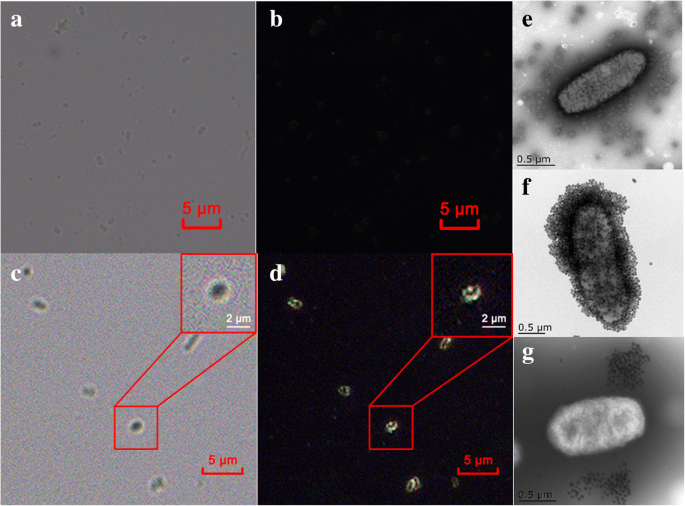


Ultrasensitive And Rapid Count Of Escherichia Coli Using Magnetic Nanoparticle Probe Under Dark Field Microscope Bmc Microbiology Full Text
Bacteria species E coli and S aureus under the microscope with different magnifications Bacteria are among the smallest, simplest and most ancient livingUnder a high magnification of 66X, this digitallycolorized, scanning electron microscopic (SEM) image depicted a growing cluster of Gramnegative, rodshaped, Escherichia coli bacteria, of the strain O157H7, which is a pathogenic strain of E coli High Resolution Click here for hiresolution image (1642 MB) Content Providers(s)A student is examining a bacterium under the microscope The E coli bacterial cell has a mass of m = 0 \rm fg (where a femtogram, \rm fg, is \rm 10^ {15}\;


Scanning Electron Microscopy The Morphology Of E Coli Cells Before Download Scientific Diagram
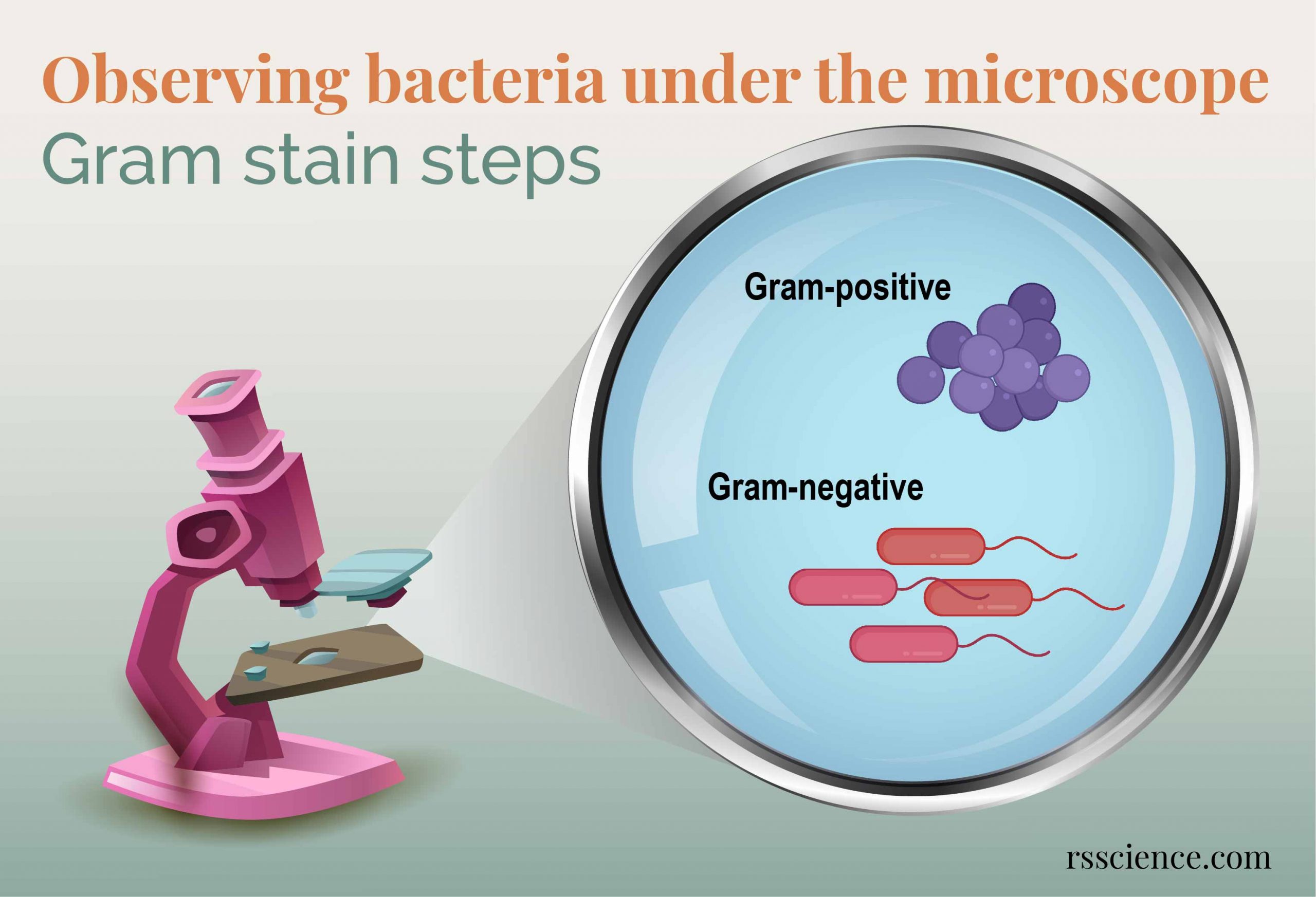


Observing Bacteria Under The Microscope Gram Stain Steps Rs Science
Optical Microscope Images Of E Coli Cells Following Gram Staining What Does An E Coli Bacteria Look Like Under A Microscope Quora Gram Stain Demonstration Slide 400x 2 A Slide Demonstrati Flickr S Epidermidis Gram Stain Escherichia Coli Free Images Public Domain ImagesEscherichia coli is shaped in the form of a rod It is a gramnegative bacterium and it contains peptidoglycan which is a thin and outer membrane Ecoli is considered a facultative thermophile, which means that it can survive under high temperatures as well as moderate temperatures It is present in most mammals particularly in the gut where it performs important functions like aiding inSpecialized structures in cells allow a cell to be able to do a certain function They allow a cell to act like a bone cell or a skin cell, for example 1 Collect data Use the microscope to observe the samples listed in the table below For each sample, estimate the cell size and


Escherichia Coli Wikipedia


Advantages Of Using Escherichia Coli As A Model Organism Andor Learning Centre Oxford Instruments
G) and is swimming at a velocity ofA student is examining a bacterium under the microscope the e coli bacterial cell has a mass of m = 150 fg (where a femtogram, fg, is 10−15g) and is swimming at a velocity of v = 900 μm/s , with an uncertainty in the velocity of 800 % e coli bacterial cells are around 1 μm ( 10−6 m) in length the student is supposed to observe the bacterium and make a drawing however, theE Coli Under Microscope Gram Stain Written By MacPride Friday, June 29, 18 Add Comment Edit E Coli Gram Stain Gram Stain Images Microbiology Wayne State


Electron Microscopy Of Bacteria Stock Photo Download Image Now Istock
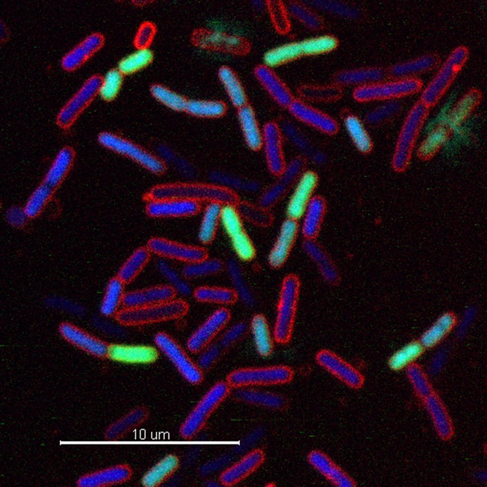


Novel Class Of Antibiotics Brings New Options
Figure 3 Spectral scan of DH5 E coli bacteria and media contents The contents of a cell growth assay plate were transferred to a Corning 3635 UV transmissible plate and scanned from 0 nm to 999 nm in 1 nm increments using a Synergy 4 MultiMode Microplate Reader with Hybrid Technology Figure 4 Effect of wavelength on AbsorbanceEscherichia coli or Ecoli is a gramnegative species of bacillus shaped bacteria that can be easily observed under a microscope, even for those with the untrained eye This bacteria has a fast growth rate, doubling every minutes, making them a common choice for bacterial related research purposesIt depedns Ecoli is a procaryotic microorganism, typically about 2 to 4 µm long, rodshaped bacterium (bacillus) with rounded ends Cells can be shorter than 2 µm or can form long filaments Ecoli is usually motile in liquid or semisolid environment with peritrichous flagella (about 6 per cell) and its surface is covered with fimbriae These structures (flagella and fimbriae) are too thin to be visualized by classical light microscopy or they don't have to be present at all under


Cross Sections Of E Coli Bacteria Producing Rsmd Viewed Under The Download Scientific Diagram
/tongue_bacteria-57b636455f9b58cdfde3ff5a.jpg)


Differences Between Bacteria And Viruses
A student is examining a bacterium under the microscope The E coli bacterial cell has a mass of m = 0300 fg (where a femtogram, fg, is 10−15g) and is swimming at a velocity of v = 900 μm/s , with an uncertainty in the velocity of 700 % E coli bacterial cells are around 1 μm ( 10−6 m) in lengthA student is examining a bacterium under the microscope The E coli bacterial cell has a mass of m = 190 fg (where a femtogram, fg, is 10−15g) and is swimming at a velocity of v = 0 μm/s , with an uncertainty in the velocity of 300 % E coli bacterial cells are around 1 μm ( 10−6 m) in lengthE Coli Clues Current TB research at HMS stems from work conducted in the bacterial model organism Escherichia coli Throughout his career, Alfred Goldberg, HMS professor of cell biology, has been studying proteases—enzymes that help break proteins down into smaller molecules or amino acids This work has led to breakthroughs in cancer


Cell Division Of E Coli With Continuous Media Flow Youtube


E Coli Cells Or Bacteria Or Virus Floating Under Microscope Stock Video Download Video Clip Now Istock
For instance, a drug can pass through membranes in order to affect a cell, and similarly, some information must pass through membrane channels to exit the cell While other cells besides E coliWhile some of these cells may be truly round shaped, others may appear elongated (ovoid) or beanshaped/kidney shaped For instance, some Neisseria cells may appear round while others are beanshaped when viewed under the microscope Tetrad bacteria Tetrad bacteria are arranged in groups of four cells Following division, the cells remainA student is examining a bacterium under the microscope The E coli bacterial cell has a mass of m = 0800fg (where a femtogram, fg, is 10−15g) and is swimming at a velocity of v = 100μm/s , with an uncertainty in the velocity of 100% E coli bacterial cells are around 1 μm ( 10−6 m) in length The student is supposed to observe the bacterium and make a drawing


551 Salmonella Bacterium Videos And Hd Footage Getty Images
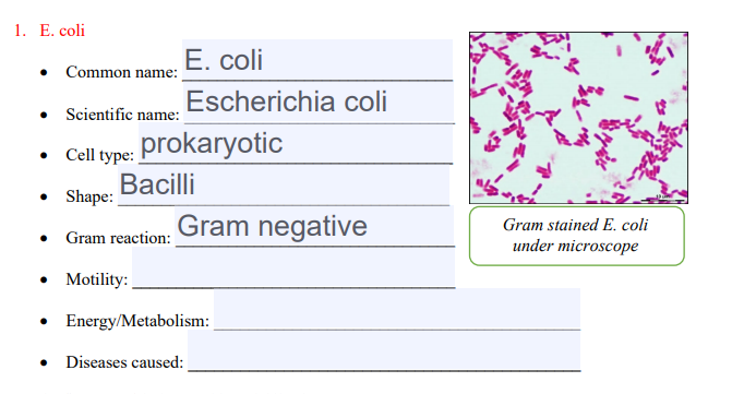


Solved 1 E Coli E Coli Common Name Escherichia Coli S Chegg Com
Place the slide on a staining rack and add a few drops of crystal violet onto the sample, gently wash with water Add a few drops of Gram iodine (mordant) for between 30 seconds and 1 minute, gently wash with water Add a few drops of alcohol (95% alcohol) for about 10 seconds, gently wash with waterE coli under the microscope Hanging drop method This technique is performed to observe the motility of the organism Under this method, the living Simple staining This method is usually performed to detect and observe E coli simply Here, the organism is stained Gram Staining After GramIt is 13 x 0407 µm in size and 06 to 07 µm in volume It is arranged singly or in pairs It is motile due to peritrichous flagella Some strains are nonmotile Some strains may be fimbriated The fimbriae are of type 1 (hemagglutinating & mannosesensitive) and are present in both motile and nonmotile strains
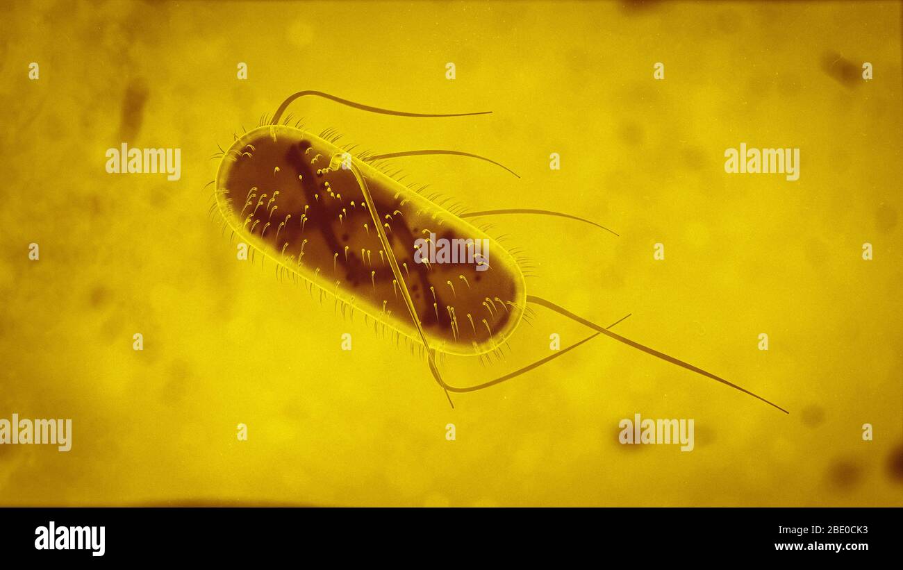


3d Escherichia Coli E Coli Cells Or Bacteria Under Microscope 3d Illustration Stock Photo Alamy
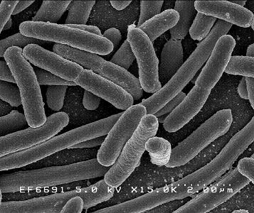


Escherichia Coli Microbewiki
2 Morphology and Staining of Escherichia Coli E coli is Gramnegative straight rod, 13 µ x 0407 µ, arranged singly or in pairs (Fig 281) It is motile by peritrichous flagellae, though some strains are nonmotile Spores are not formed Capsules and fimbriae are found in some strains 3 Cultural Characteristics of Escherichia ColiSpecialized structures in cells allow a cell to be able to do a certain function They allow a cell to act like a bone cell or a skin cell, for example 1 Collect data Use the microscope to observe the samples listed in the table below For each sample, estimate the cell size andWhich bacteria look similar to E coli under 100X optical microscope?


Escherichia Coli Bacteria E Coli Stock Footage Video 100 Royalty Free Shutterstock


Medical Concept Viral Or Bacterial Diseases Coronavirus Sars Stock Photo Picture And Royalty Free Image Image
Bio 112 Abstract Escherichia coli, Bacillus subtilis and Staphylococcus epidermidis were analyzed for this lab activity to determine their Gram Stain After the multilayered Gram Stain procedure each bacteria were classified as Grampositive or Gramnegative depending on their cell walls staining color The results showed that E coli stained pink and classified as GramnegativeBacteria S aureus, Lactobacillus sp, Bacillus anthracis, E coli, etc Conclusion Yeast is a eukaryotic organism while bacteria are prokaryotes Both yeast and bacteria are unicellular organisms with a cell wall Yeast contains a nucleus and membranebound organelles but, bacteria lack a nucleus or membranebound organellesEscherichia coli (E coli) bacteria normally live in the intestines of people and animals Most E coli are harmless and actually are an important part of a healthy human intestinal tract However, some E coli are pathogenic, meaning they can cause illness, either diarrhea or illness outside of the intestinal tract The types of E coli that can cause diarrhea can be transmitted through


E Coli Ancient Aliens Health Science Electron Microscope


Aph162 Report 1
Instead, their genetic material floats uncovered, localized to a region called the nucleoidThe WHO estimates up to 700,000 patients died in of multidrug resistant bacterial infections globally in 16 This rise of multidrug resistant (MDR) bacteriExamples of Bacteria Under the Microscope Escherichia coli Escherichia coli (Ecoli) is a common gramnegative bacterial species that is often one of the first ones to be observed by students Most strains of Ecoli are harmless to humans, but some are pathogens and are responsible for gastrointestinal infections They are a bacillus shaped bacteria that has a very fast growth (they can double every minutes), which is one of the main reasons they are used in research
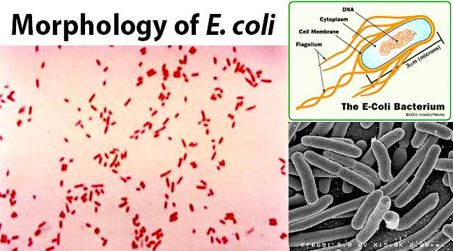


Escherichia Coli E Coli An Overview Microbe Notes


Interrogating The Escherichia Coli Cell Cycle By Cell Dimension Perturbations Pnas
Escherichia coli bacteria (E coli) under microscope, magnification 1000X D By Dimarion Stock footage ID Video clip length 0021 FPS 2997 Aspect ratio 169 Standard footage license HD $79 1280 × 7 MOVA student is examining a bacterium under the microscope The E coli bacterial cell has a mass of = 0500 (where a femtogram,fg , is 1015 g) and is swimming at a velocity of v = 900 um/s, with an uncertainty in the velocity of 800 % E coli bacterial cells are around 1 um (106 m ) in length The student is supposed to observe the bacterium and make a drawingE coli bacteria looks like small rods under compound microscope at 1000 magnification power It is gram negative bacterium which appears pink when stained by a staining technique called as Gram staining which is a differential staining technique
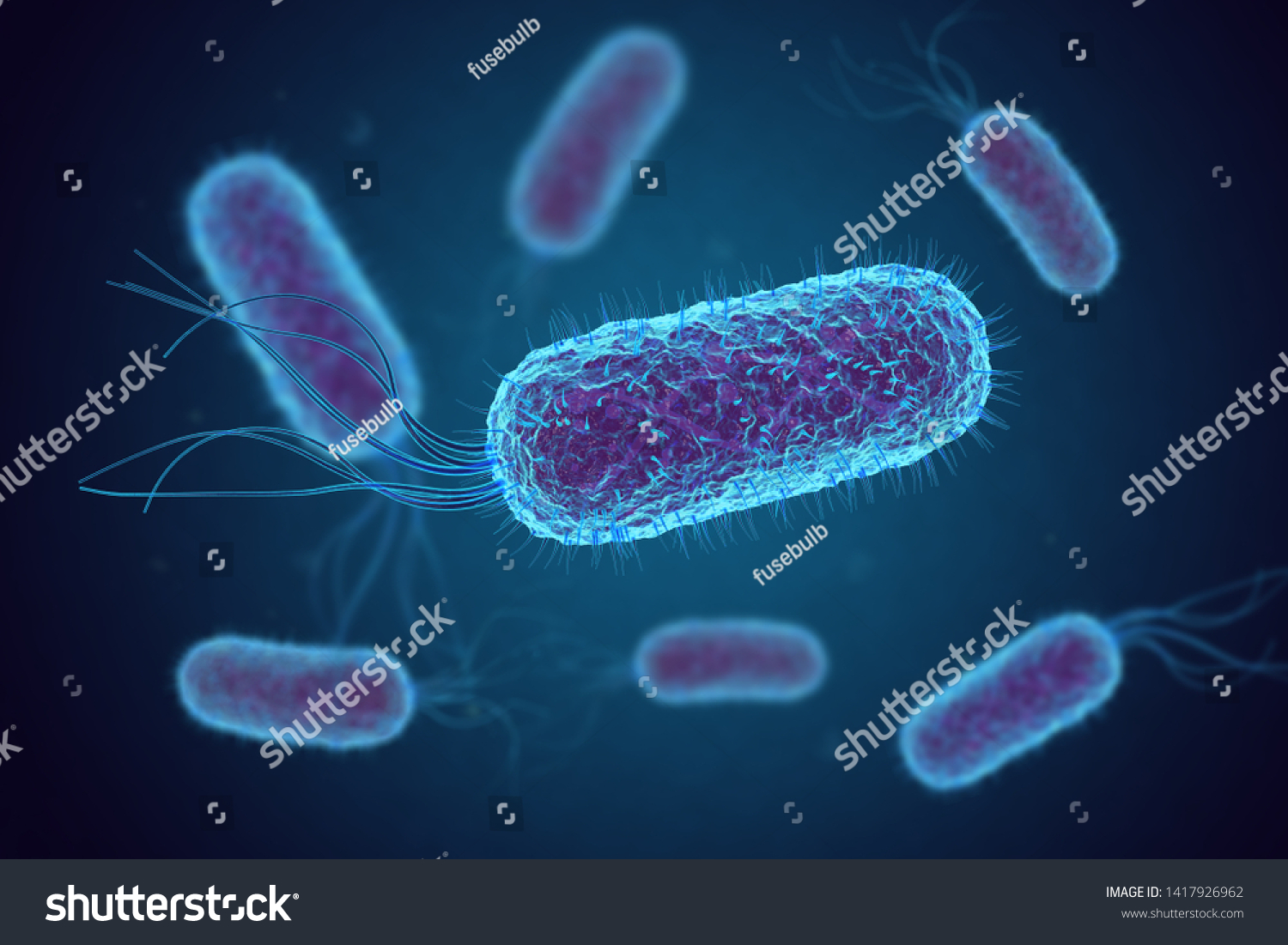


Escherichia Coli E Coli Cells Bacteria Stock Illustration


Ryu Lab Movies
They began to study cell division of cells under a microscope to determine how operons worked They began testing different chemicals on E coli to determine the number of operons presentEscherichia coli bacteria (E coli) under microscope, magnification 1000X D By Dimarion Stock footage ID Video clip length 0021 FPS 2997 Aspect ratio 169 Standard footage license HD $79 1280 × 7 MOVWhen viewed under the microscope, Gramnegative E Coli will appear pink in color The absence of this (of purple color) is indicative of Grampositive bacteria and the absence of Gramnegative E Coli Escherichia coli under 10х90х magnification using fuchsine as a dye by ElNokko (Own work) CC BYSA 40 (http//creativecommonsorg/licenses/bysa/40), via Wikimedia Commons
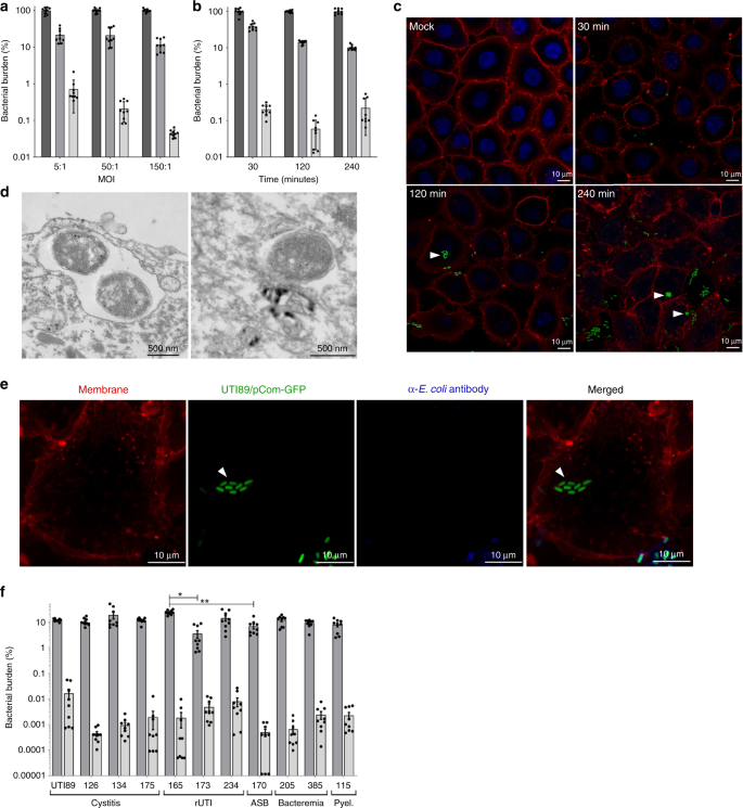


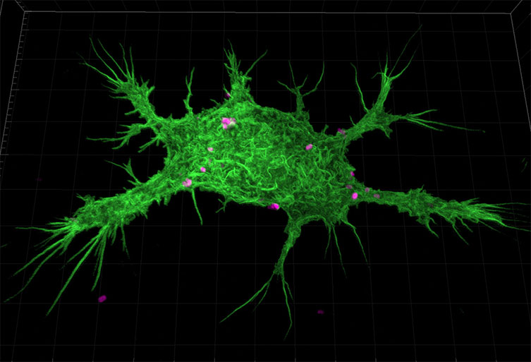
Invasion Of Vaginal Epithelial Cells By Uropathogenic Escherichia Coli Nature Communications
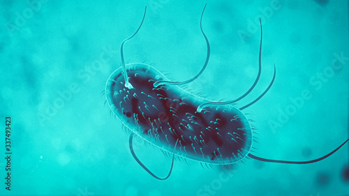


3d Escherichia Coli E Coli Cells Or Bacteria Under Microscope 3d Illustration Buy This Stock Illustration And Explore Similar Illustrations At Adobe Stock Adobe Stock
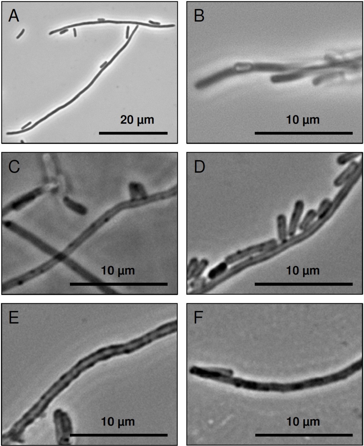


Development Of Functionalised Polyelectrolyte Capsules Using Filamentous Escherichia Coli Cells Microbial Cell Factories Full Text



Various Bacteria Cells In Microscope Streptococcus Pneumonia P Stock Photo Image By C Maxxyustas


Escherichia Coli Wikipedia


E Coli Bacteria Microscopic Organism Indicates Fecal Contamination
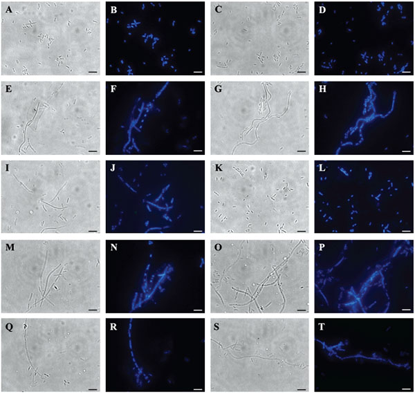


Expression Of Phi11 Gp07 Causes Filamentation In Escherichia Coli
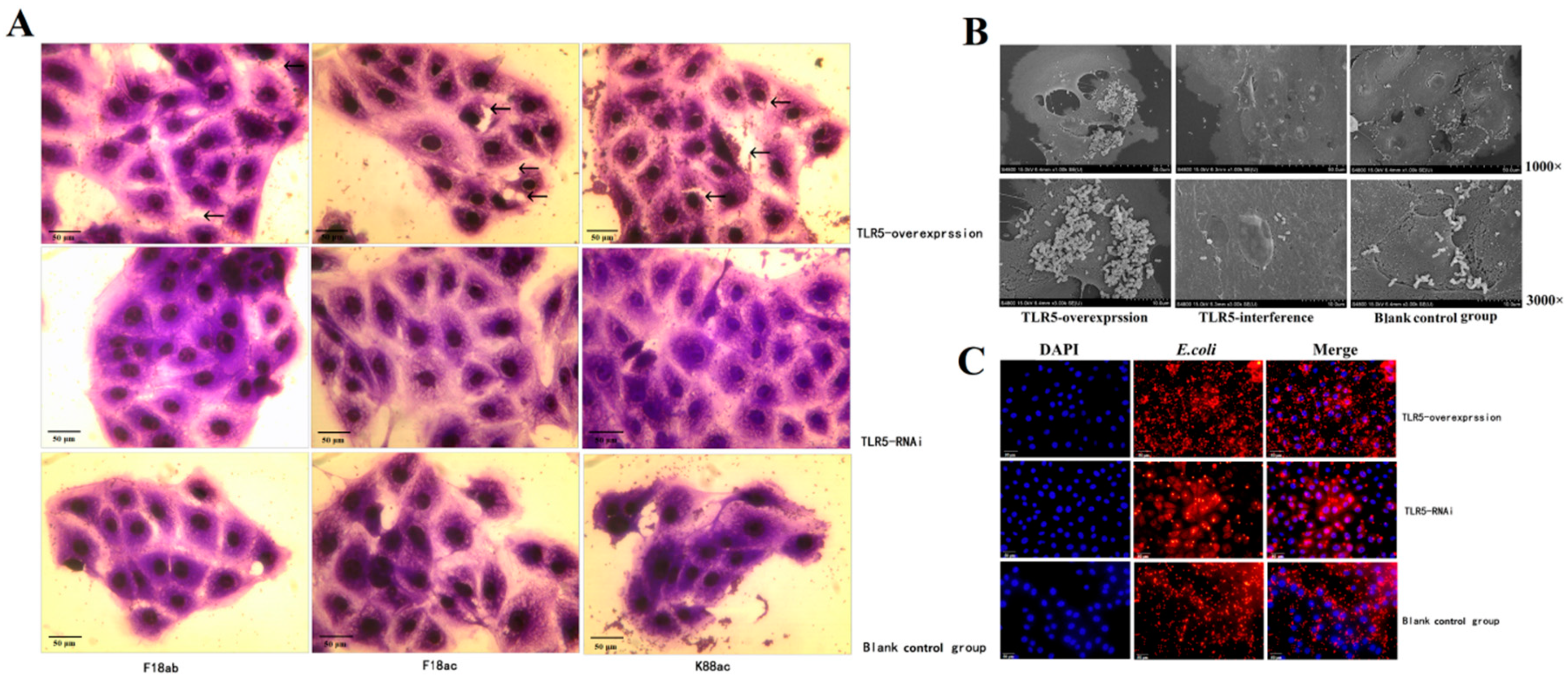


Animals Free Full Text Regulation And Molecular Mechanism Of Tlr5 On Resistance To Escherichia Coli F18 In Weaned Piglets


Insight Into Why E Coli Sickness Levels Vary
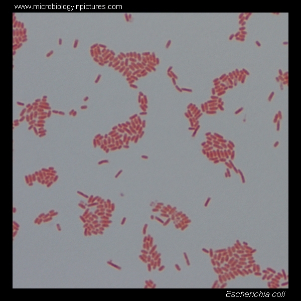


E Coli Gram Stain And Cell Morphology E Coli Micrograph Appearance Under The Microscope E Coli Cell Morphology E Coli Microscopic Picture


Striatisporolide A A Butenolide Metabolite From Athyrium Multidentatum Doll Ching As A Potential Antibacterial Agent


High Precision Characterization Of Individual E Coli Cell Morphology By Scanning Flow Cytometry Konokhova 13 Cytometry Part A Wiley Online Library
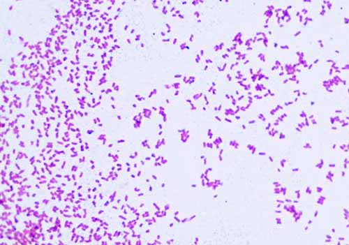


Gram Negative Bacteria Images Photos Of Escherichia Coli Salmonella Enterobacter


Magnification Bioninja


Introductory Chapter The Versatile Escherichia Coli Intechopen


Small Things Considered E Coli Cells Face Facs And Get Back Into Shape


Pdf Nanoscale Imaging Of E Coli Cells By Expansion Microscopy Semantic Scholar
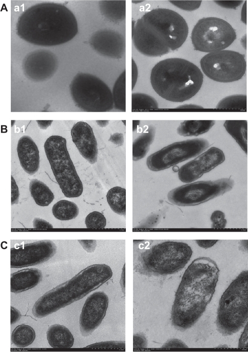


Morphology And Structure Of Bacterial Cells Under Trans Open I


Antibacterial Activity Of A Terpineol May Induce Morphostructural Alterations In Escherichia Coli
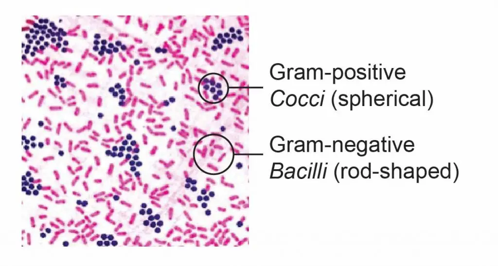


Observing Bacteria Under The Microscope Gram Stain Steps Rs Science
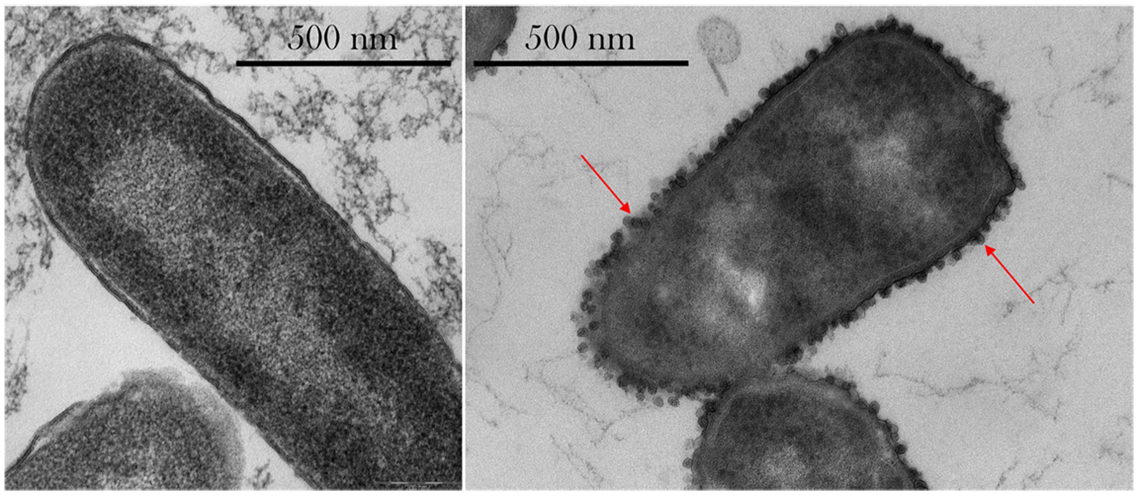


Frontiers Phenotypic Changes Exhibited By E Coli Cultured In Space Microbiology


E Coli Bateria Microscopic Electron Microscope Images Biology Poster
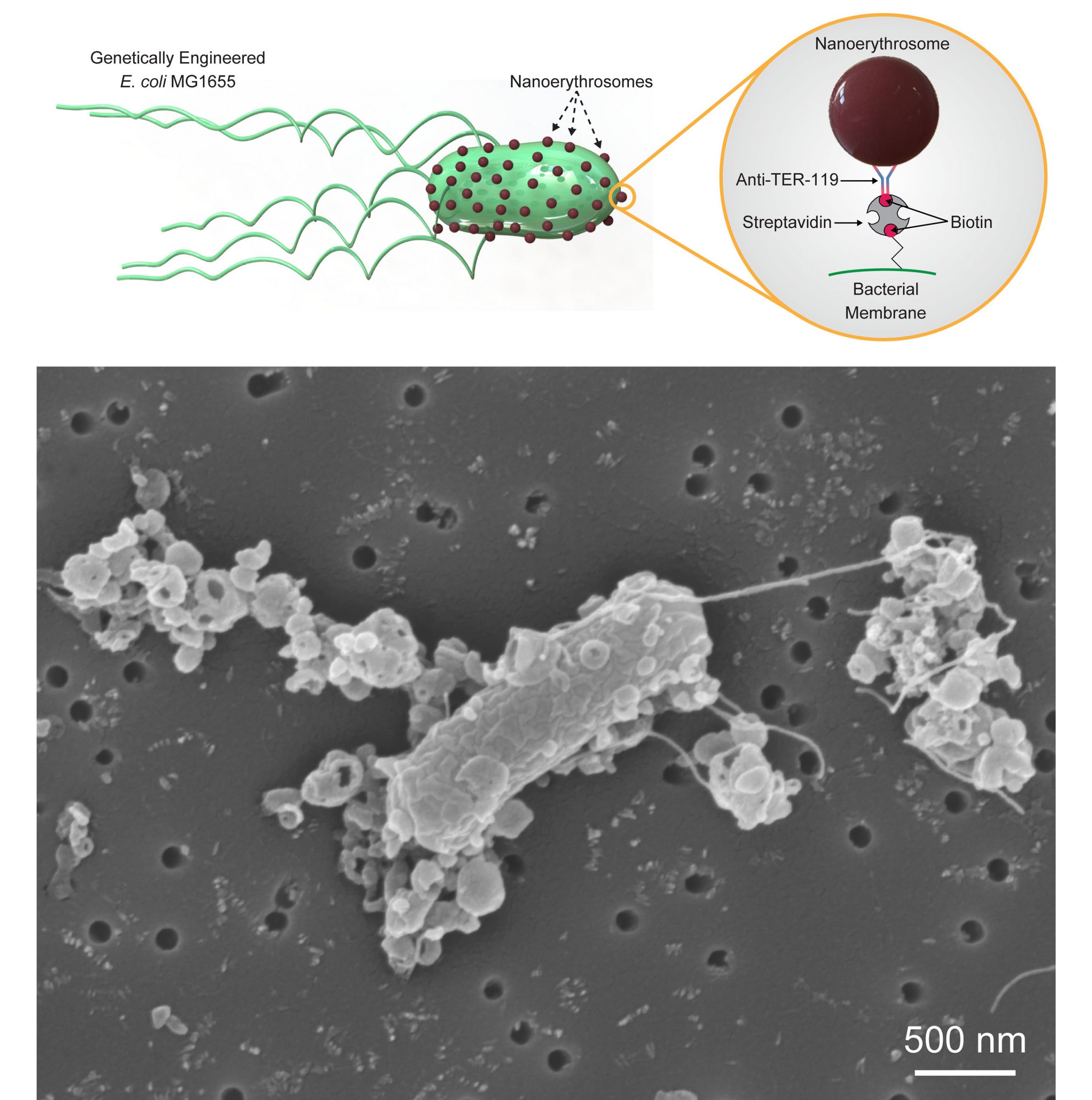


Personalized Microrobots Swim Through Biological Barriers Deliver Drugs To Cells Aip Publishing Llc


Q Tbn And9gcqy Atzkf0po8c8x1yoohusnpjlpu6pnvquhzmi5dmxlldlhkom Usqp Cau


Morphology Of E Coli Cells Under Microscope At 100 Magnification Download Scientific Diagram


Tech Tip Imaging Bacteria Using Agarose Pads Biotium
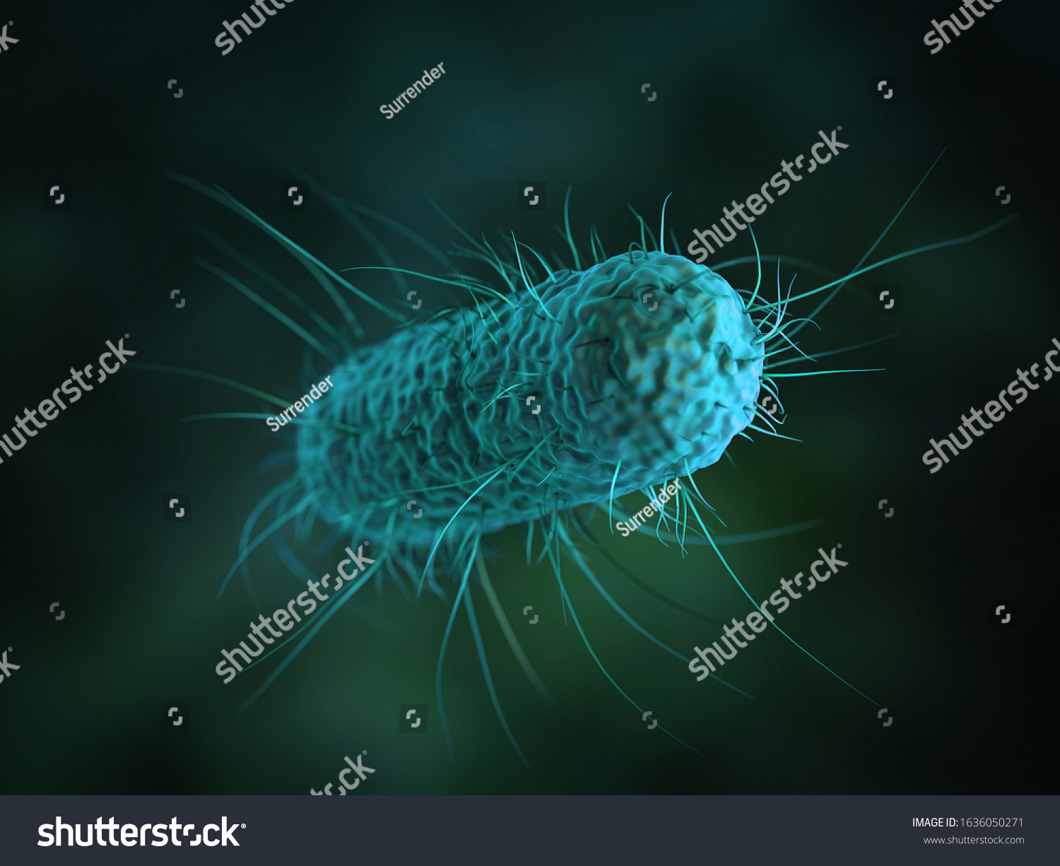


Close 3d Microscopic Bacteria Cells Escherichia Stock Illustration


Morphologies Of Recombinant E Coli Cells As Observed By Phase Contrast Download Scientific Diagram


Optical Microscope Images Of E Coli Cells Following Gram Staining A Download Scientific Diagram
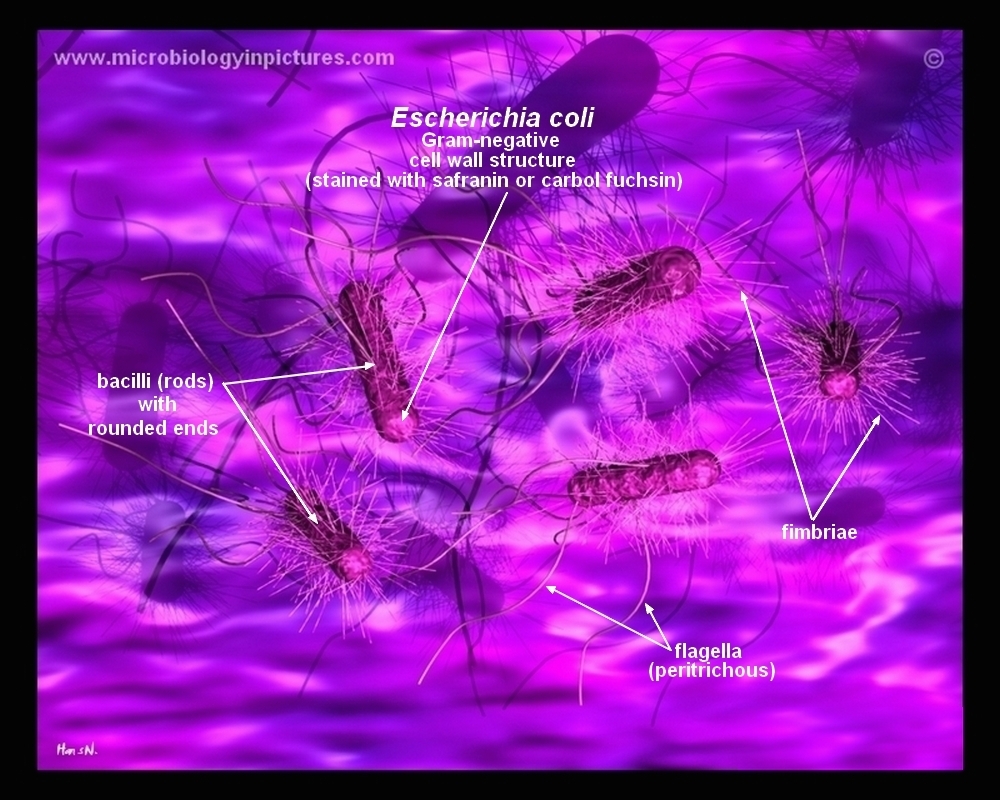


How E Coli Bacteria Look Like
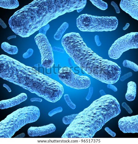


Shutterstock Puzzlepix


Direct Visualization Of Escherichia Coli Chemotaxis Receptor Arrays Using Cryo Electron Microscopy Pnas


Biol 230 Lab Manual Gram Stain Of E Coli


E Coli Bacteria Light Microscopy Stock Video Clip K004 9042 Science Photo Library


Morphological And Physiological Changes Induced By High Hydrostatic Pressure In Exponential And Stationary Phase Cells Of Escherichia Coli Relationship With Cell Death Applied And Environmental Microbiology


Microscopic Studies Of Various Organisms
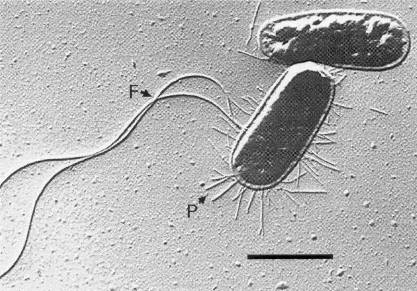


Escherichia Coli 07 Igem Org


Microscopic Studies Of Various Organisms


Bacteria Capsules And Slime Layers Britannica
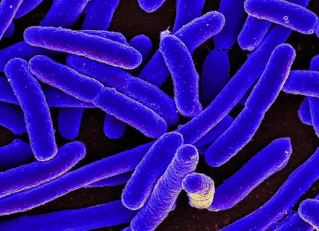


E Coli Under The Microscope Types Techniques Gram Stain Hanging Drop Method


Bacteria Growth


Escherichia Coli Light Microscopy
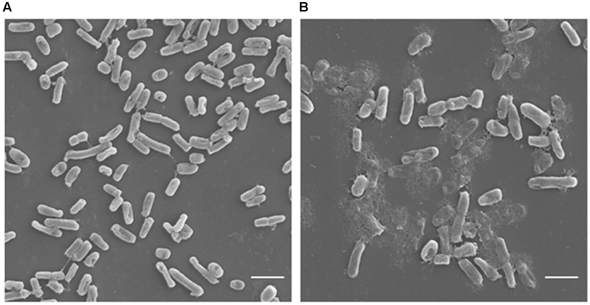


Frontiers Antimicrobial Potential Of Carvacrol Against Uropathogenic Escherichia Coli Via Membrane Disruption Depolarization And Reactive Oxygen Species Generation Microbiology
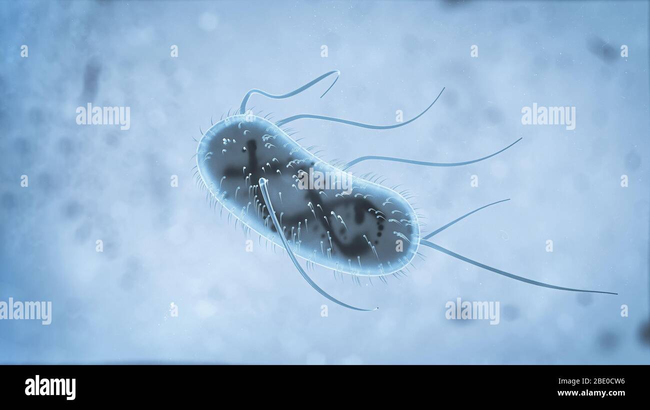


3d Escherichia Coli E Coli Cells Or Bacteria Under Microscope 3d Illustration Stock Photo Alamy
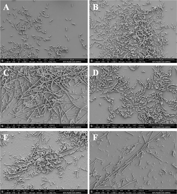


Expression Of Phi11 Gp07 Causes Filamentation In Escherichia Coli Fulltext


E Coli Cell Arrangement Page 1 Line 17qq Com


Photomicrographs


2 2 Prokaryotic Cells Bioninja


A Microscale Method Of Protein Extraction From Bacteria Interaction Of Escherichia Coli With Cationic Microparticles Sciencedirect
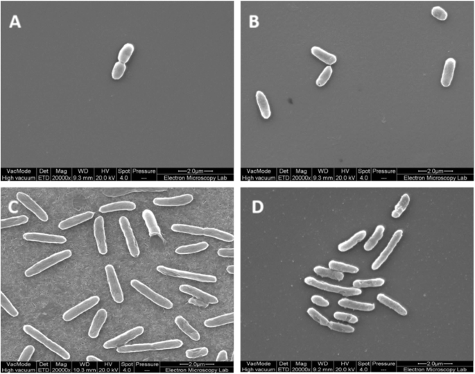


Thymol Tolerance In Escherichia Coli Induces Morphological Metabolic And Genetic Changes Bmc Microbiology Full Text


Electron Microscope Used To Highlight Cranberry Activity


Microscope Imaging Of Methylene Blue Stained E Coli Cells Harbouring Download Scientific Diagram


Markus Covert How To Build A Computer Model Of A Cell Stanford School Of Engineering


Team Lethbridge Results 11 Igem Org


Bacterial Flagella Disrupt Host Cell Membranes And Interact With Cytoskeletal Components Biorxiv


Introductory Chapter The Versatile Escherichia Coli Intechopen


Bacteria Under The Microscope E Coli And S Aureus Youtube


19 599 Bacterium Videos And Hd Footage Getty Images


Pathogenic E Coli


Escherichia Coli Colony Morphology And Microscopic Appearance Basic Characteristic And Tests For Identification Of E Coli Bacteria Images Of Escherichia Coli Antibiotic Treatment Of E Coli Infections
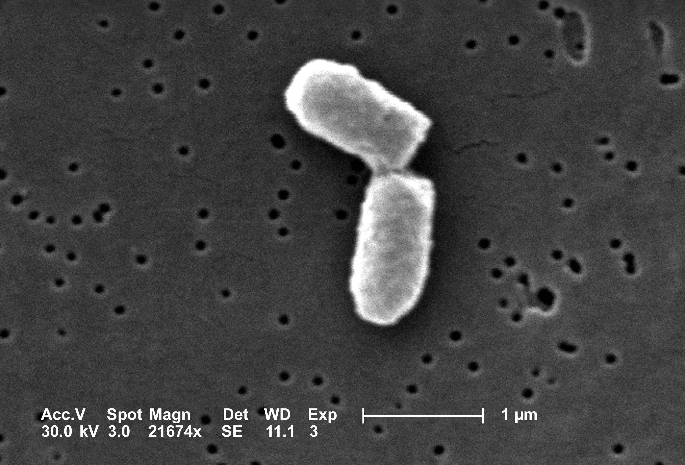


How Long Would It Take Bacteria To Eat The Earth By Niko Mccarty Medium


Genomes Of Two Popular Research Strains Of E Coli Sequenced


How Big Is An E Coli Cell And What Is Its Mass


Indole Prevents Escherichia Coli Cell Division By Modulating Membrane Potential Sciencedirect
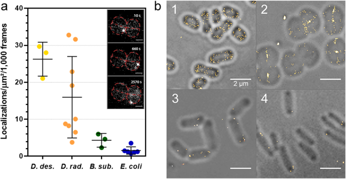


Bacterial Cell Wall Nanoimaging By Autoblinking Microscopy Scientific Reports


E Coli Under A Scanning Electron Microscope Image Eurekalert Science News


The Unique Tropism Of Mycobacterium Leprae To The Nasal Epithelial Cells Can Be Explained By The Mammalian Cell Entry Protein 1a


Q Tbn And9gcr1gxralqmge Lwsjjzsouihvmyiv0ajl7a5fu8ma5voxpjs64l Usqp Cau


How E Coli Cells Work In The Human Gut Uconn Today


A Structural Study Of Escherichia Coli Cells Using An In Situ Liquid Chamber Tem Technology
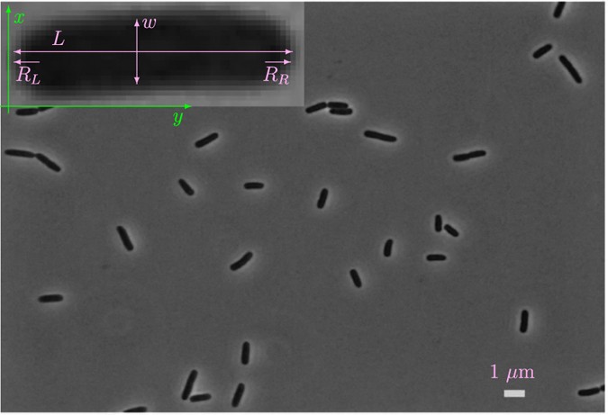


Evidence Of Multi Domain Morphological Structures In Living Escherichia Coli Scientific Reports


Q Tbn And9gcrxsv6valktvydnaiqm6 Yoim4dbn 9td0atqk2azvfczn73wcq Usqp Cau


What Does An E Coli Bacteria Look Like Under A Microscope Quora


Gram Stain Wikipedia
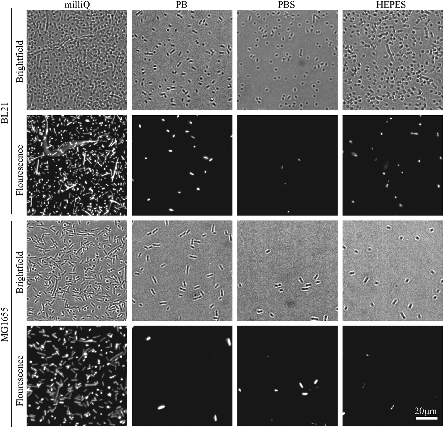


Imaging Live Bacteria At The Nanoscale Comparison Of Immobilisation Strategies Analyst Rsc Publishing Doi 10 1039 C9and


Q Tbn And9gcqkye60ou Johpr02n Mbv1fferrjpdh Lnct7ymdf5qhyia1ld Usqp Cau


コメント
コメントを投稿